Direct Immunofluorescence (DIF) Quiz
DIF is a key diagnostic tool in dermatology used to detect the deposition of immunoreactants (IgG, IgA, IgM, C3, fibrin) in autoimmune skin diseases. Master your diagnostic skills with these clinical cases!
Progress: 0/9
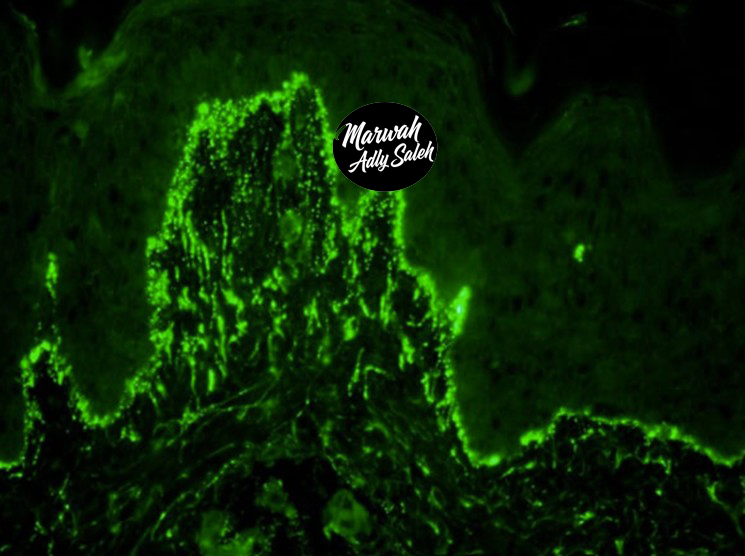
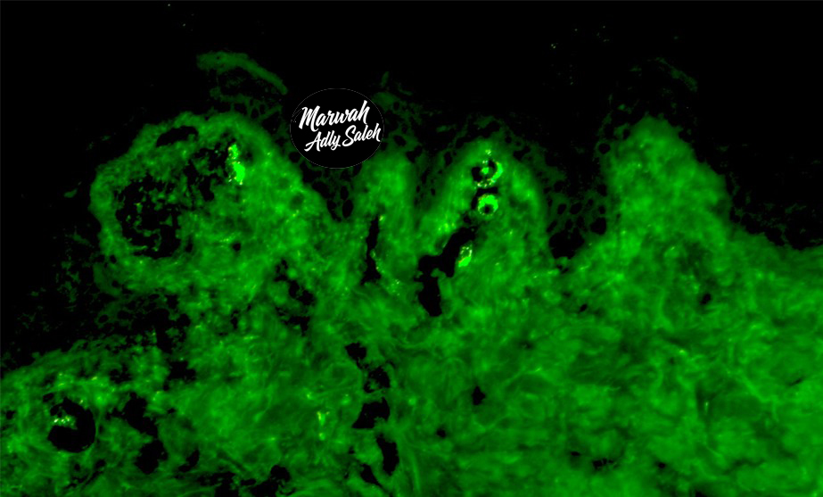
Case 1
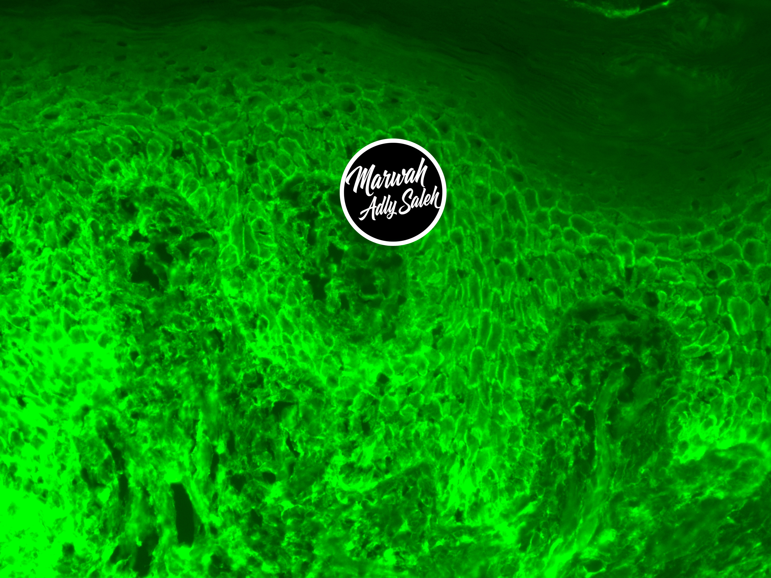
Clinical History:
A 45-year-old patient presents with painful oral erosions and flaccid skin blisters. A skin biopsy was taken from perilesional skin of an erosion. The biopsy was stained with IgG, IgM, IgA, and C3.
What is your diagnosis?
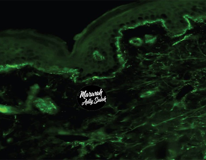
Case 2
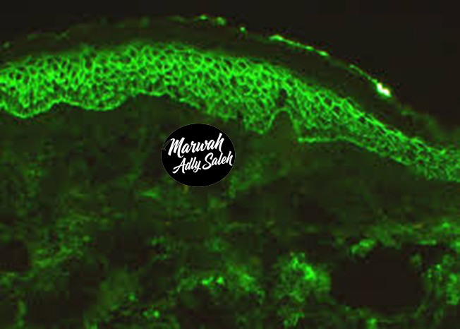
Clinical History:
A 50-year-old patient with facial erythema and blisters. A skin biopsy was taken from perilesional skin of an erosion.
What is your diagnosis?
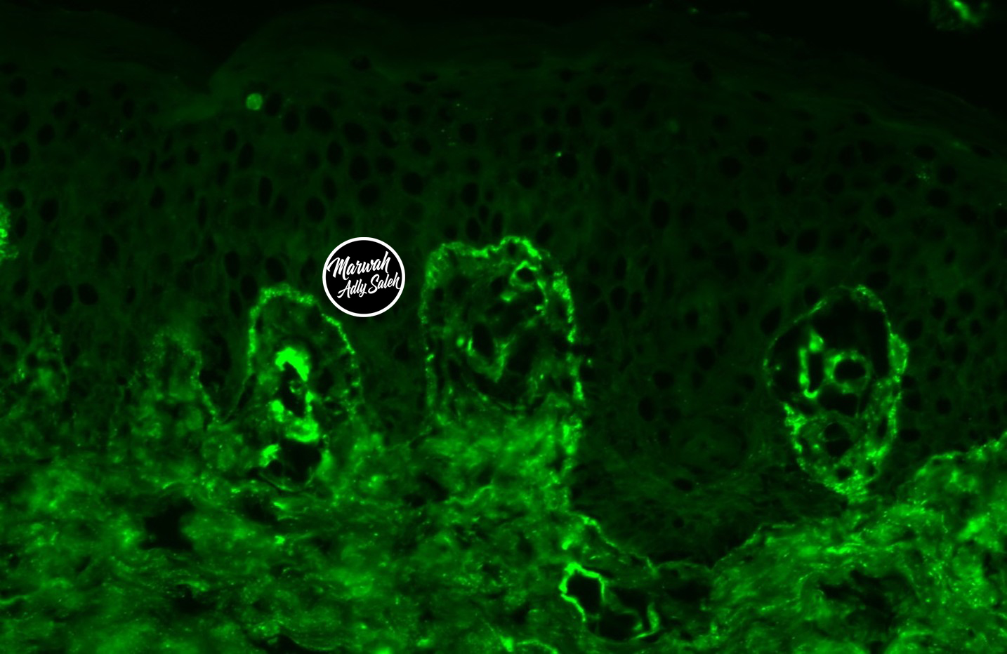
Case 3
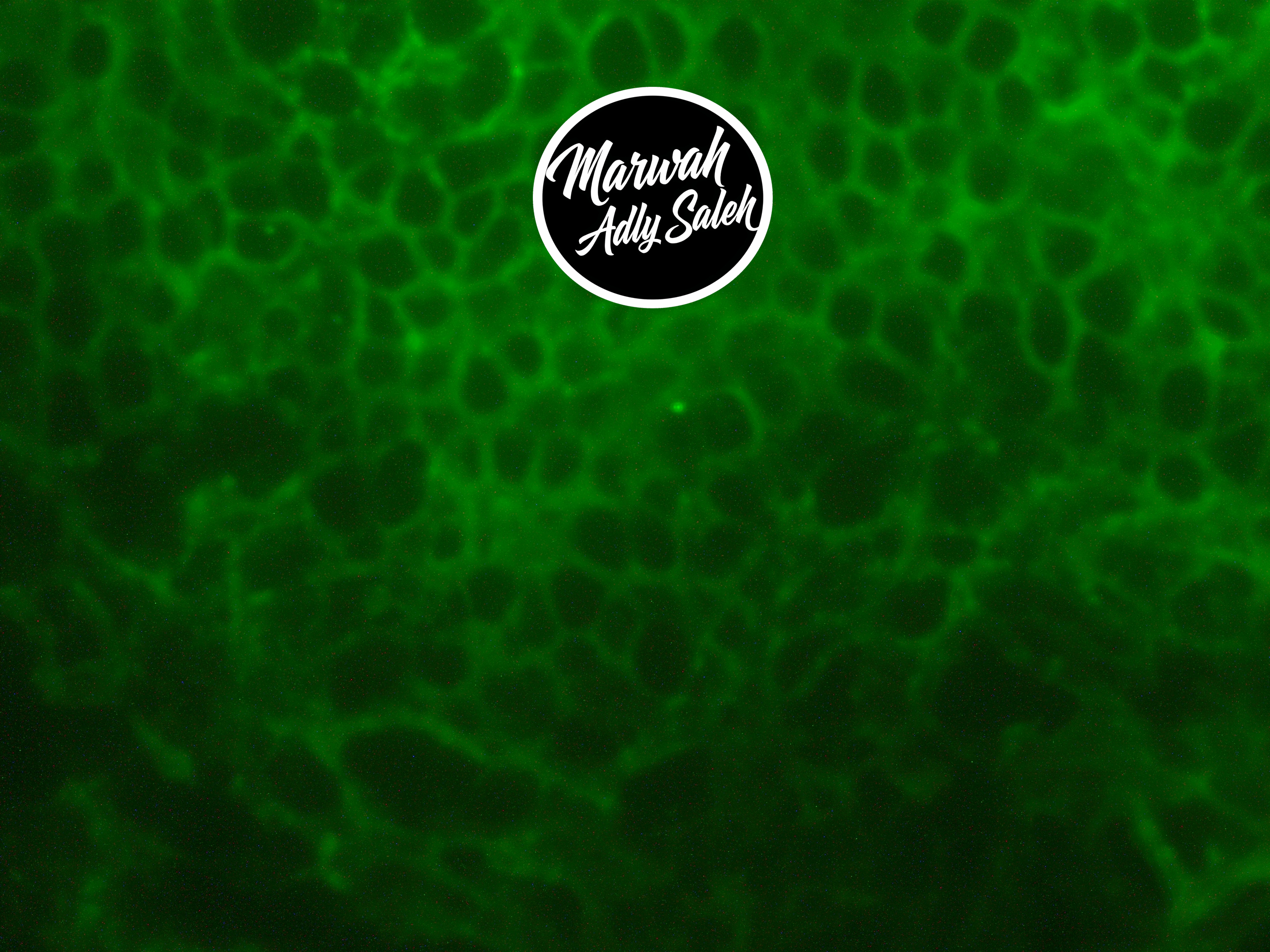
Clinical History:
A 65-year-old patient with severe mucosal erosions and recent diagnosis of lymphoma. A mucosal biopsy was taken from perilesional mucosa of an erosion.
What is your diagnosis?
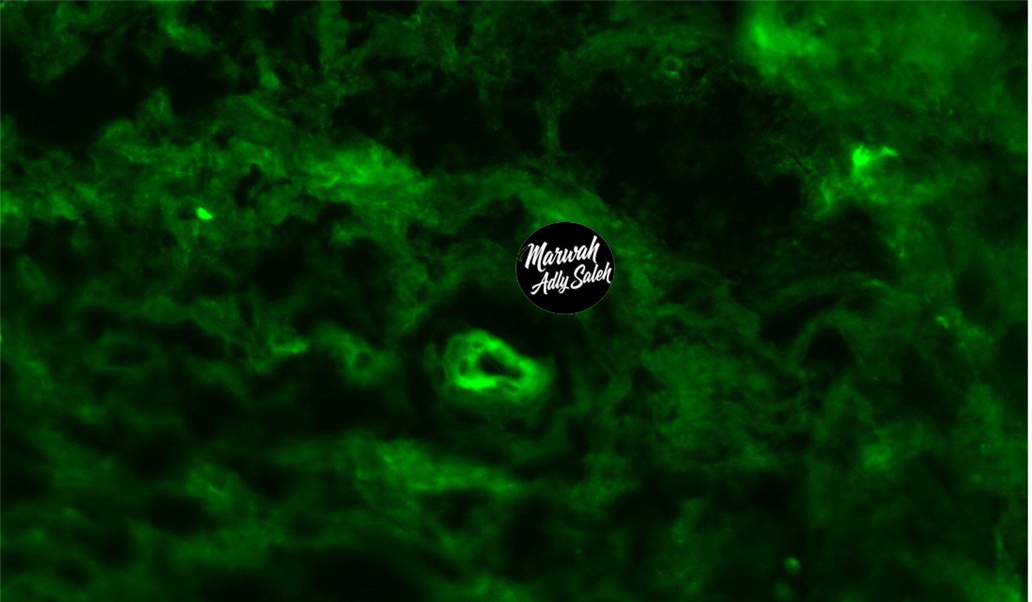
Case 4
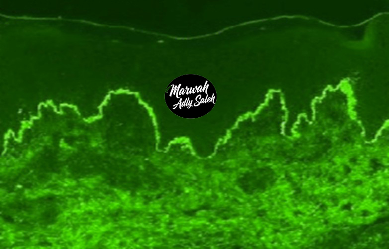
Clinical History:
A 70-old patient with tense blisters on Lower limbs. A skin biopsy was taken from perilesional skin of a blister.
What is your diagnosis?
Case 5
Clinical History:
A 35-year-old patient with intensely pruritic papules and vesicles on extensor surfaces. Skin biopsy was taken from a papule on the extensor surface.
What is your diagnosis?
Case 6
Clinical History:
A 25-year-old woman with butterfly rash and joint pain. A skin biopsy was taken from an erythematous plaque on sun-exposed skin.
What is your diagnosis?
Case 7
Clinical History:
A 25-year-old woman with butterfly rash and joint pain. A skin biopsy was taken from an erythematous plaque on sun-exposed skin.
What is your diagnosis?
Case 8
Clinical History:
A 55-year-old patient with palpable purpura on lower extremities. A skin biopsy was taken from a palpable purpura lesion.
What is your diagnosis?
Case 9
Clinical History:
A 8-old child with palpable purpura on lower extremities, abdominal pain, and hematuria. A skin biopsy was taken from a palpable purpura lesion.